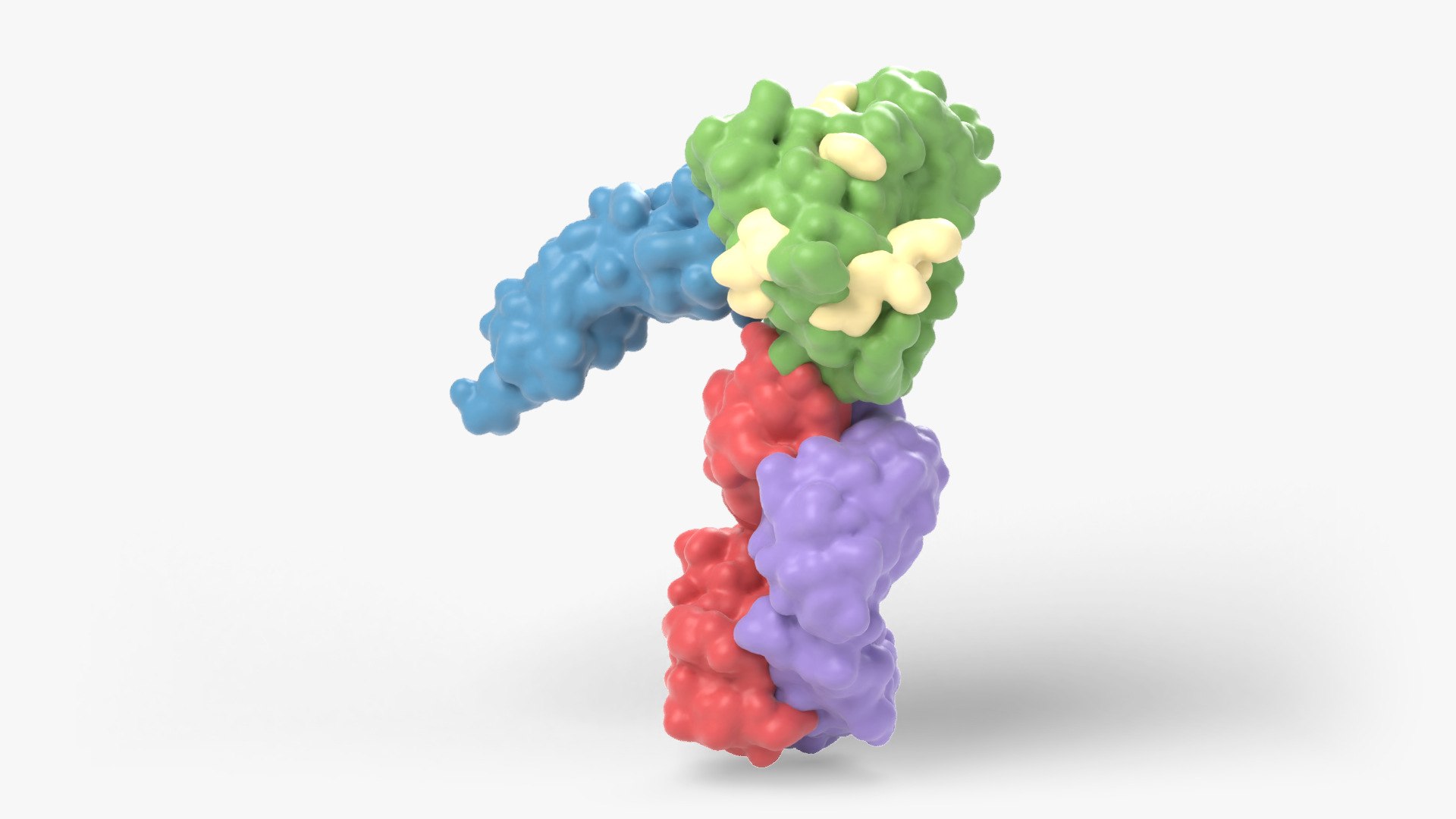
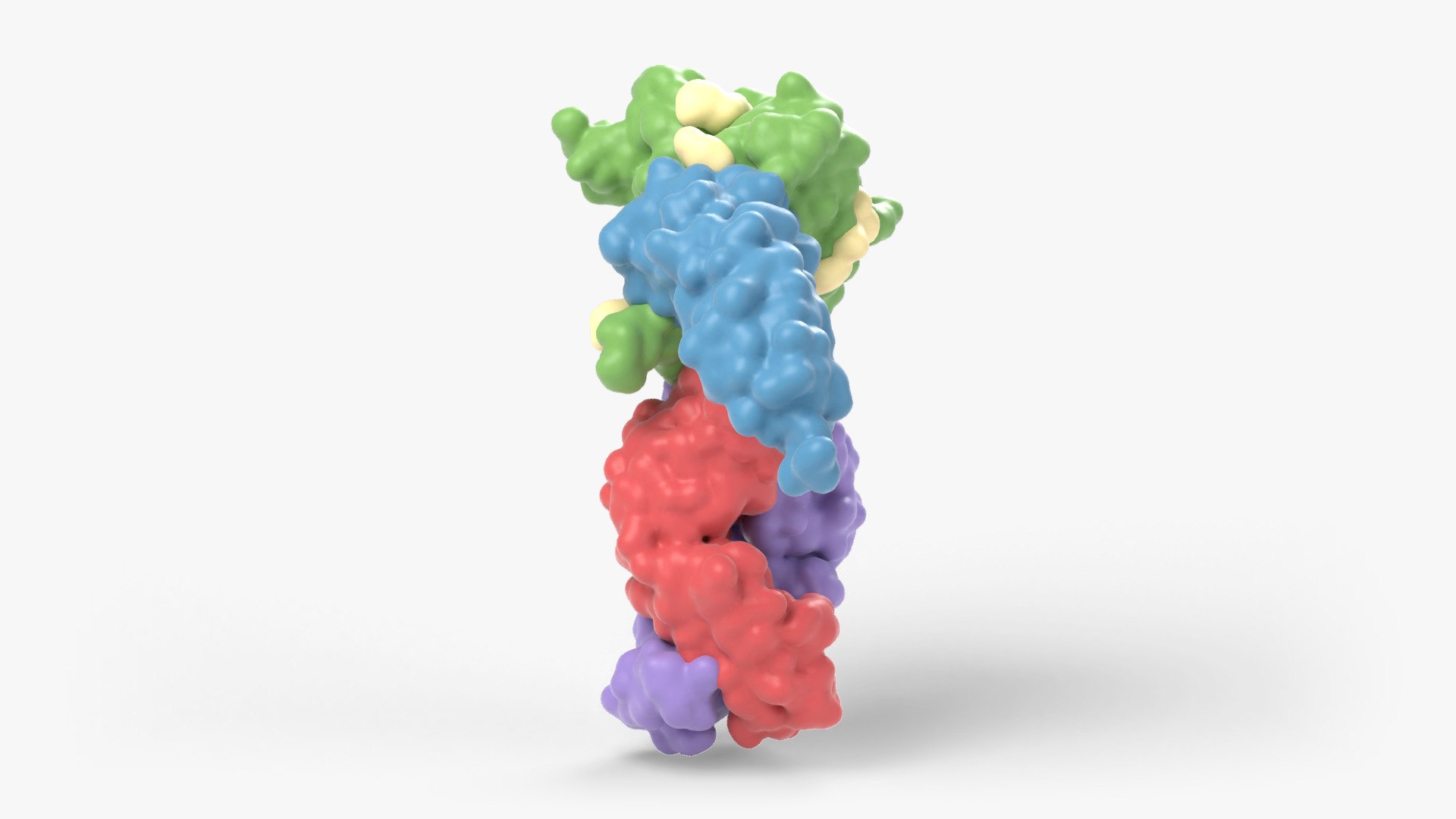
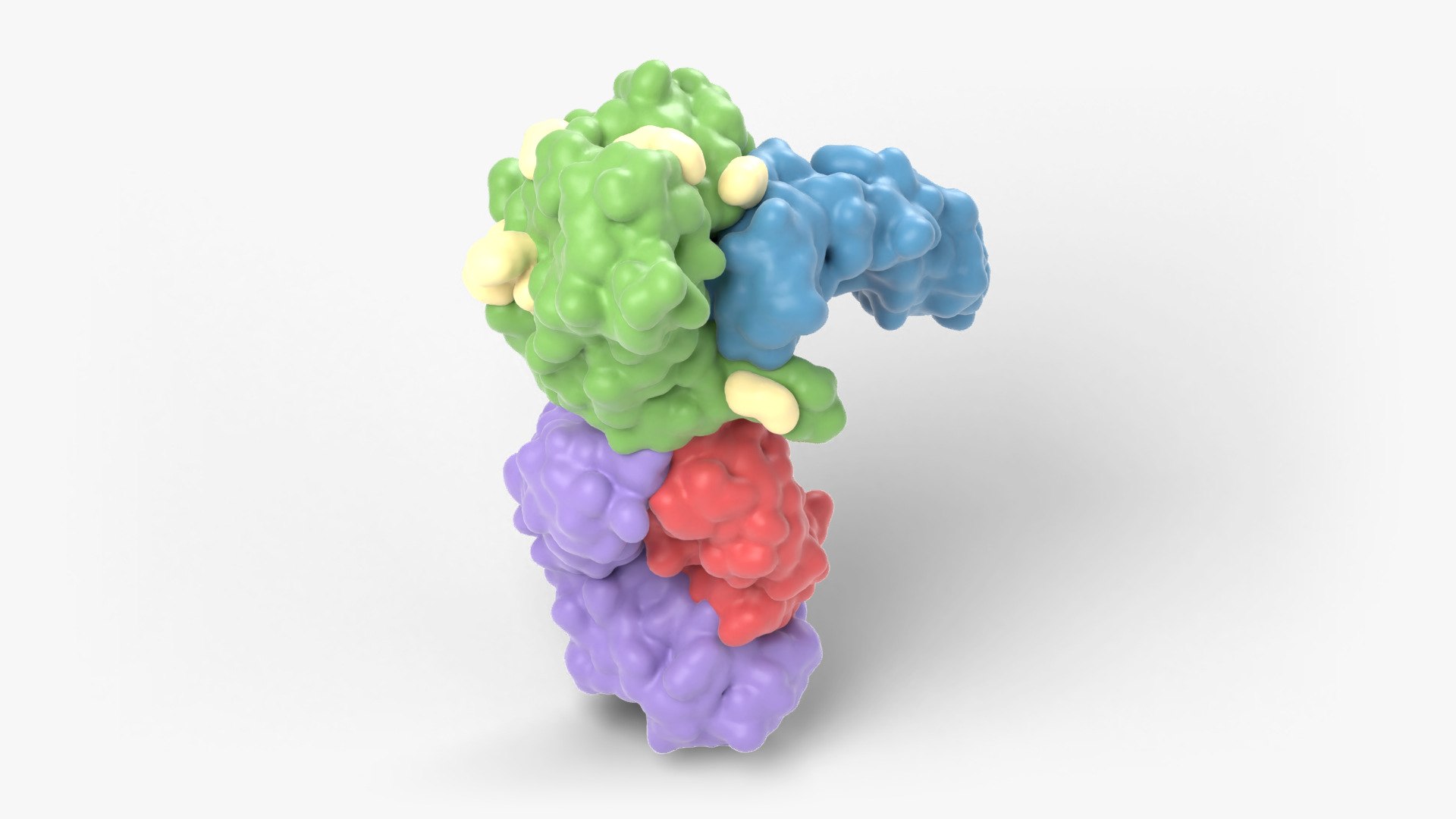
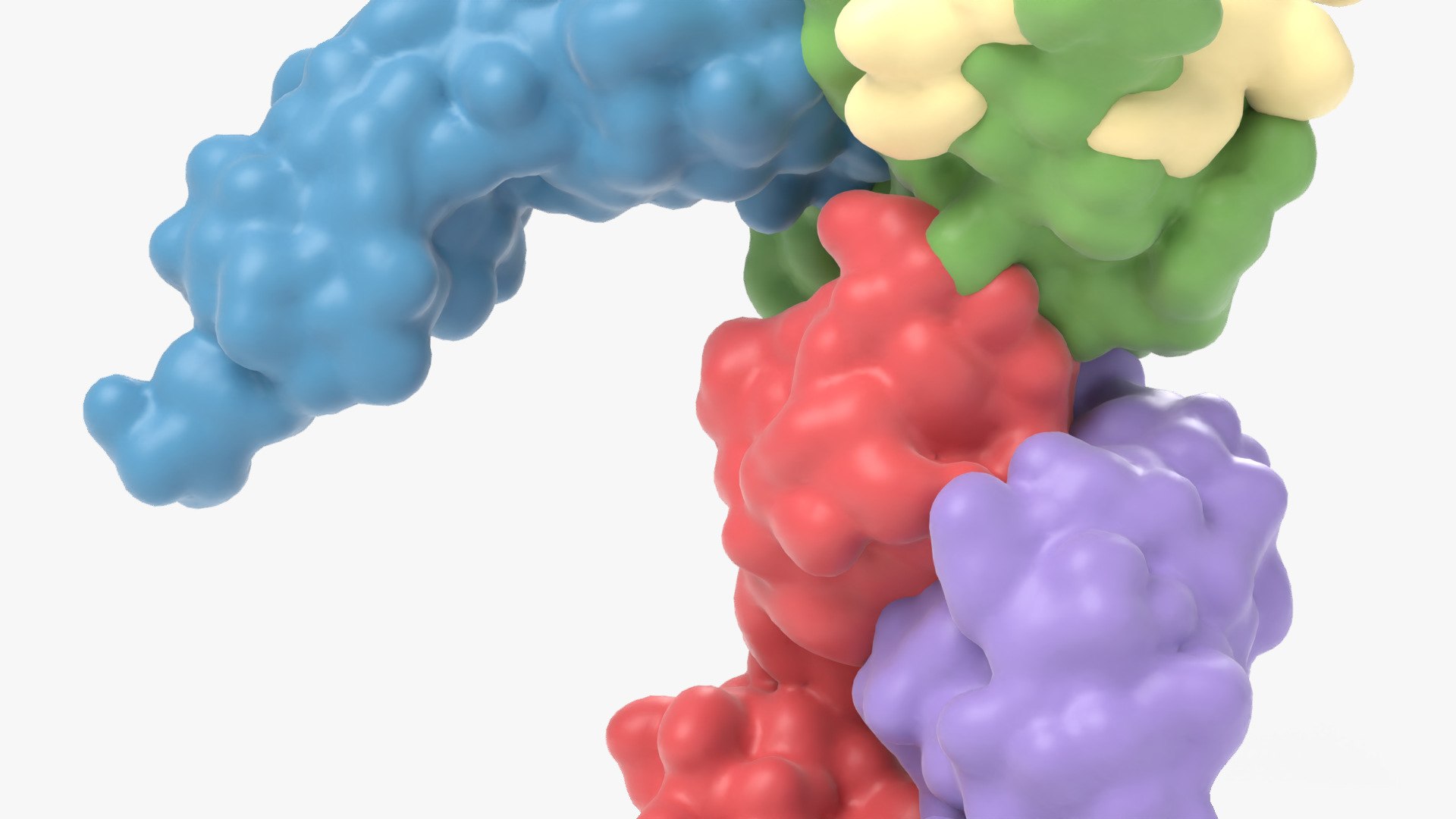
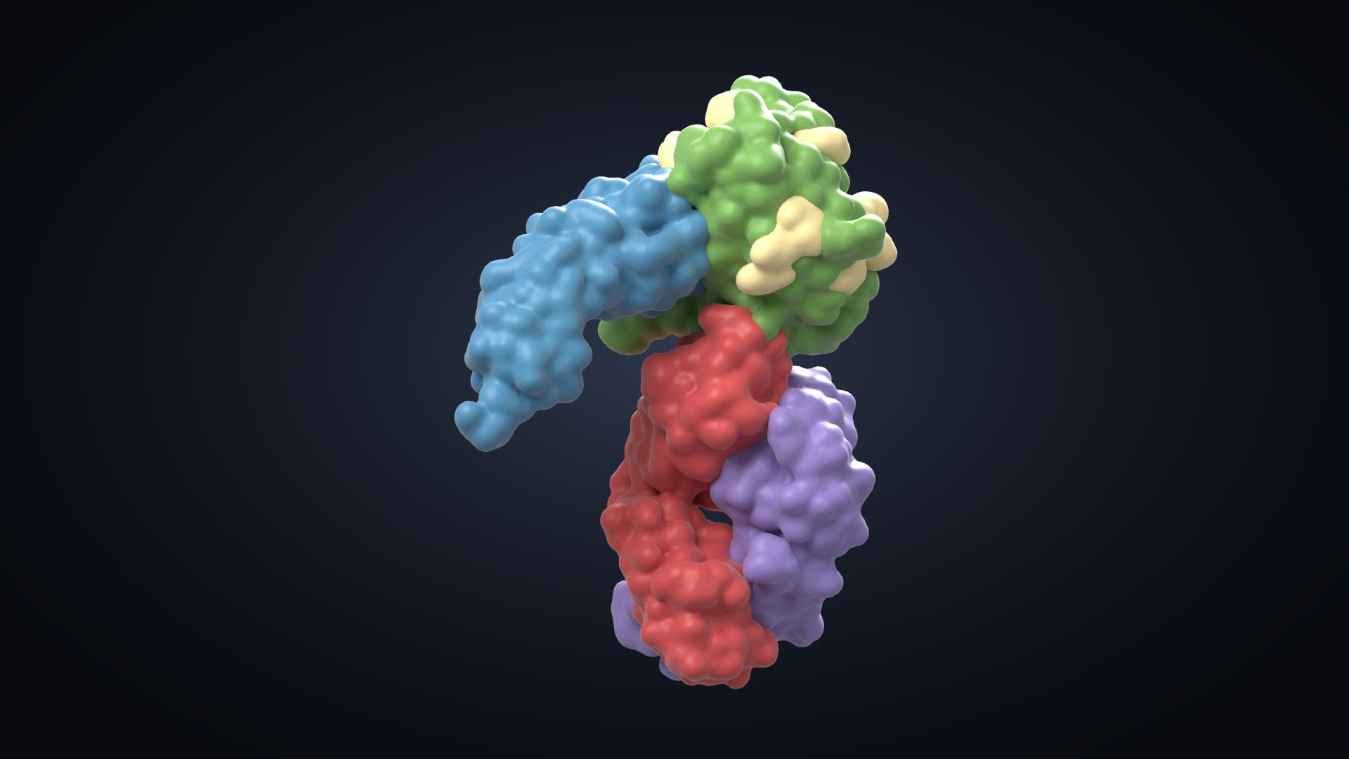
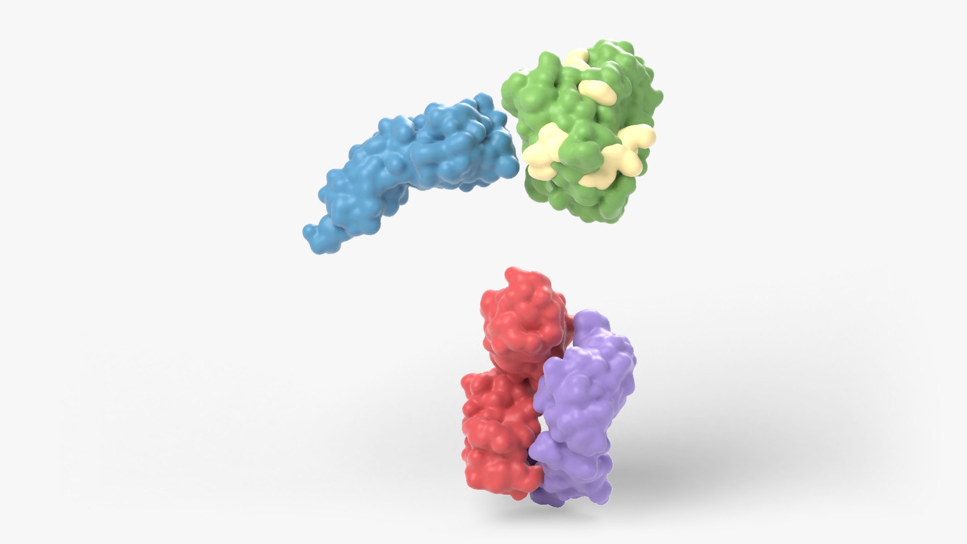
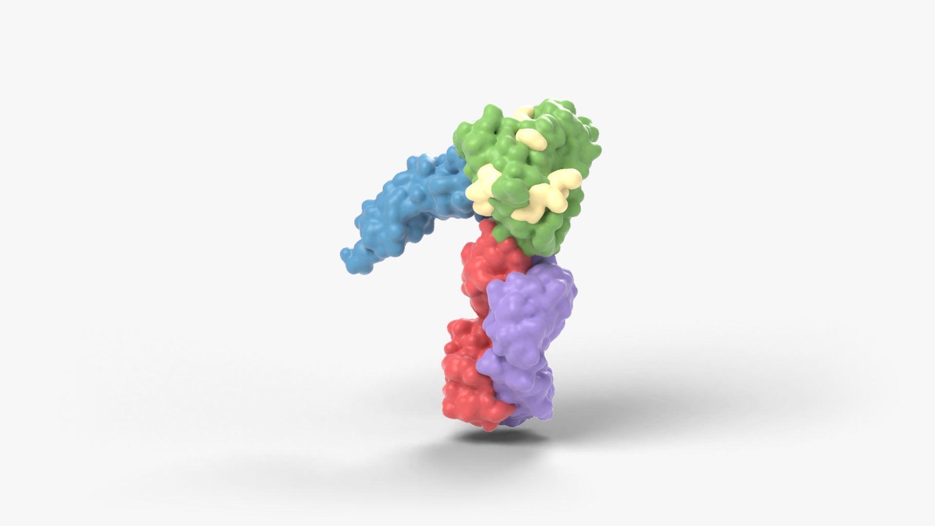
Core of the HIV-1 envelope protein gp120 bound to the human CD4 receptor and to the Fab of a neutralising antibody. The structure shows how the virus attaches to the cells it infects.
Scientifically accurate model built directly from the experimental atomic coordinates of Protein Data Bank entry 1GC1 (X-ray crystallography, 2.50 angstrom resolution), biological assembly 1. Source organism: Human immunodeficiency virus 1, Homo sapiens. Only atoms that were resolved in the experiment are represented; nothing has been added or invented.
MODEL
- Molecular surface of every chain as a separate, named object (4 chains, 4 distinct components)
- Surface glycans (sugar chains) as separate objects
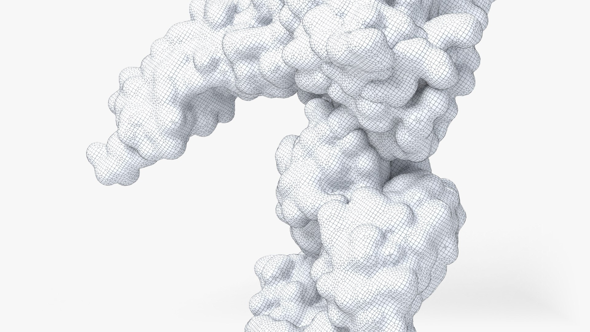
- 88,320 polygons, 88,336 vertices; all quads
- Every surface mesh is closed and manifold (no holes, no loose or overlapping vertices)
- One material per component (Principled BSDF); colours are easy to change. No textures needed
- Scale: 1 unit = 1 nanometer. Model centred at the origin, transforms clean, objects grouped under one parent
FORMATS
- Blender 4.0 (.blend), native, with the studio lighting and camera used for the previews (Cycles)
- FBX (.fbx)
- OBJ (.obj + .mtl)
- glTF 2.0 binary (.glb)
- USD (.usdc)
- STL (.stl), geometry only
Preview images were rendered in Blender Cycles. The wireframe image shows the actual mesh.
Structure reference: PDB 1GC1; Kwong et al., Nature, 1998 (doi:10.1038/31405). PDB structure data are in the public domain (CC0 1.0).